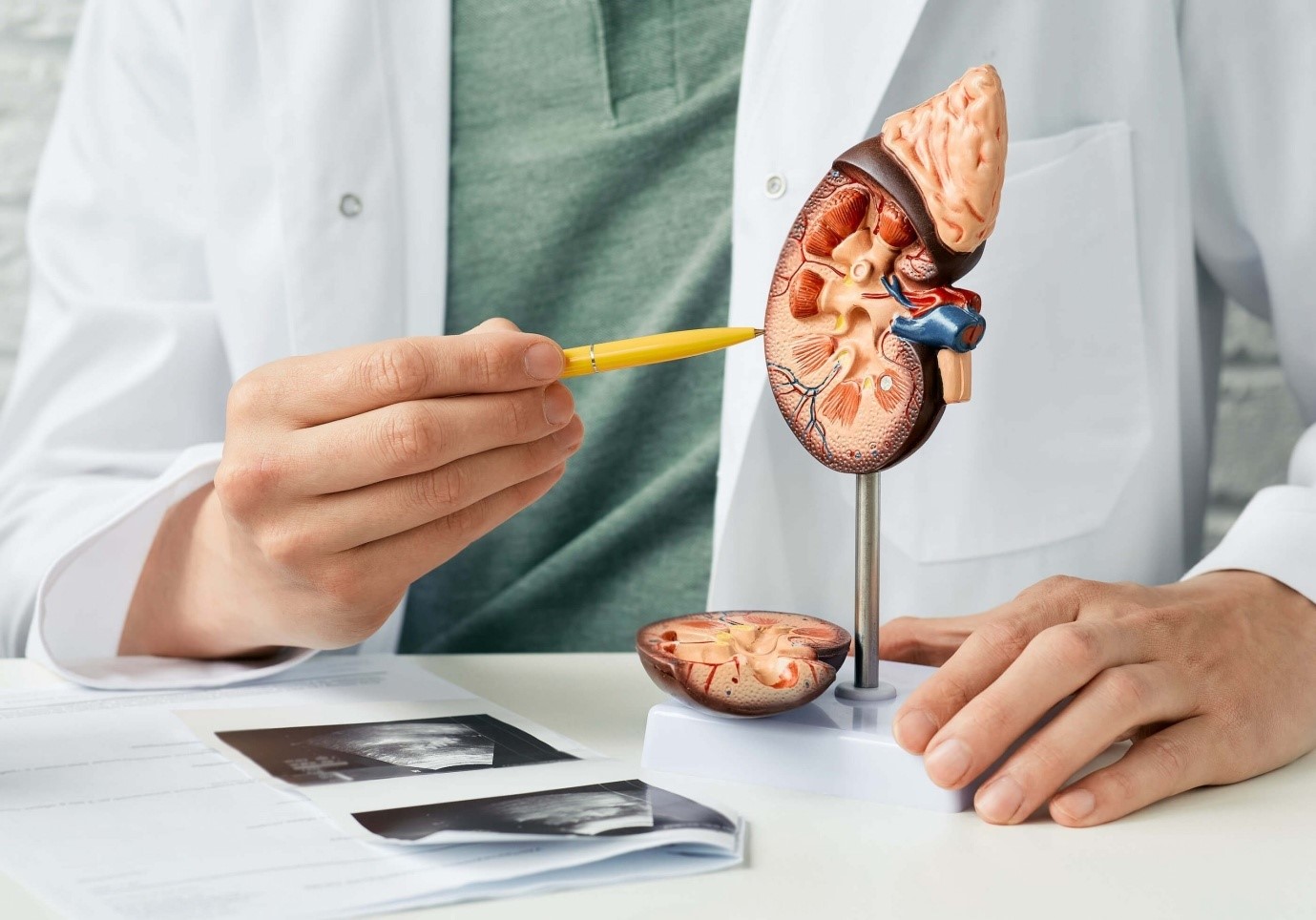
Kidney Stones

Overview
There are several types of kidney stones, each formed from different substances. The most common types include:
- Calcium stones: These are the most prevalent type and can be composed of calcium oxalate or calcium phosphate.
- Struvite stones: Formed in response to urinary tract infections, these stones may grow quickly and become large, causing blockages.
- Uric acid stones: Result from high levels of uric acid in the urine, often associated with conditions like gout or certain metabolic disorders.
- Cystine stones: Form due to a hereditary disorder called cystinuria, causing the kidney to excrete excessive amounts of certain amino acids.
After your first attack of kidney stones, there is an 80-90% chance of recurrence within the next 10 years. Knowing which kind of stone you are prone to can aid in choosing a diet that reduces or prevents recurrences.
For all stone types, the best lifestyle change to reduce recurrences is to restrict sodium and drink 3 liters or more of fluids per day.
Symptoms and Causes
Kidney stones, also known as renal calculi, are hard deposits that form in the kidneys and can cause a range of symptoms. The symptoms of kidney stones can vary depending on the size and location of the stone. Common symptoms include:
- pain in the side of your tummy (abdomen) or groin – men may have pain in their testicles
- a high temperature
- feeling sweaty
- severe pain that comes and goes
- feeling sick or vomiting
- blood in urine
- urine infection
Diagnosis And Treatment
Diagnosis:
- Medical History and Physical Examination:
Your doctor will inquire about your medical history, including any family history of kidney stones.
A physical examination may be conducted to check for signs of pain or tenderness.
- Imaging Tests:
CT Scan: This is one of the most common methods to identify kidney stones and determine their size and location.
Ultrasound: This non-invasive imaging technique may be used to visualize the kidneys and urinary tract.
X-rays: In some cases, a series of X-rays known as an intravenous pyelogram (IVP) may be performed after injecting a contrast dye.
- Urinalysis:
Examination of a urine sample can help identify crystals or substances that promote stone formation.
Treatment:
- Pain Management:
Pain relief is often a priority, and medications such as nonsteroidal anti-inflammatory drugs (NSAIDs) or opioids may be prescribed.
- Medical Expulsion Therapy (MET):
Medications like alpha-blockers may be given to help relax the muscles of the ureter, making it easier for the stone to pass.
- Hydration:
Drinking plenty of water is essential to help flush out the stone. Increased fluid intake can prevent further stone formation.
- Dietary Changes:
Depending on the type of kidney stone, your doctor may recommend dietary modifications, such as reducing salt or oxalate intake.
- Observation:
Small stones may pass on their own with supportive measures. Your doctor may recommend observation while monitoring symptoms.
- Extracorporeal Shock Wave Lithotripsy (ESWL):
This non-invasive procedure uses shock waves to break the kidney stones into smaller pieces, making them easier to pass.
- Ureteroscopy:
A thin, flexible tube with a camera is used to visualize and remove stones in the ureter or kidney.
- Percutaneous Nephrolithotomy (PCNL):
For larger stones, a surgical procedure may be required to remove or break them up.